
Introduction
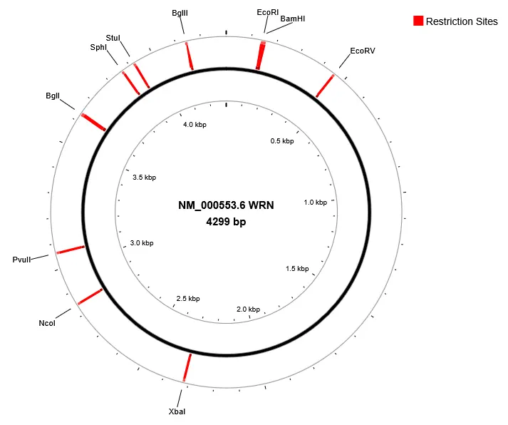
Human WRN gene (figure 1) encodes a member of the RecQ subfamily of DNA helicase proteins. This protein contains a N-terminal 3' to 5' exonuclease domain, an ATP-dependent helicase domain and RecQ helicase domain in its central region, and a C-terminal HRDC (helicase RNase D C-terminal) domain and nuclear localization signal. For MSI-H cancer cells, inhibitors that target their WRN proteins, may induce synthetic lethality. As a result, irreversible DNA damage occurs and thereby cause selective cell death.
| Experimental Specifications | |||
|---|---|---|---|
| Technique | Reagent | Instrument | Supporting |
General Procedure
I. Construction of GST-WRN500-946 plasmid vector
- Synthesize DNA corresponding to the amino acid sequence for WRN fragment of interest. In this case, amino acid (aa) residues 500-946 to form the WRN500-946 RecQ helicase protein. This specific helicase domain has been implicated to function for DNA helicase unwinding activity
- Identify the nucleotide sequence and corresponding amino acid sequence from NCBI BLAST for WRN500-946
- 2.1. The full-length Human WRN RecQ like helicase (WRN), mRNA is provided as reference nucleotide ID NM_000553.6 corresponding to 7,575 nucleotides (nt) that includes the bifunctional 3'-5' exonuclease/ATP-dependent helicase WRN at the 24 - 4,539 nt position encoding for aa sequence NP_000544.2 [Consensus CDS Protein Set: CCDS6082.1] translates to 1432 aa sequence
- 2.2 The entire 1432 aa sequence corresponding to NP_000544.2 is the following:
- MSEKKLETTAQQRKCPEWMNVQNKRCAVEERKACVRKSVFEDDLPFLEFTGSIVYSYDASDCSFLSEDISMSLSDGDVVGFDMEWPPLYNRGKLGKVALIQLCVSESKCYLFHVSSMSVFPQGLKMLLENKAVKKAGVGIEGDQWKLLRDFDIKLKNFVELTDVANKKLKCTETWSLNSLVKHLLGKQLLKDKSIRCSNWSKFPLTEDQKLYAATDAYAGFIIYRNLEILDDTVQRFAINKEEEILLSDMNKQLTSISEEVMDLAKHLPHAFSKLENPRRVSILLKDISENLYSLRRMIIGSTNIETELRPSNNLNLLSFEDSTTGGVQQKQIREHEVLIHVEDETWDPTLDHLAKHDGEDVLGNKVERKEDGFEDGVEDNKLKENMERACLMSLDITEHELQILEQQSQEEYLSDIAYKSTEHLSPNDNENDTSYVIESDEDLEMEMLKHLSPNDNENDTSYVIESDEDLEMEMLKSLENLNSGTVEPTHSKCLKMERNLGLPTKEEEEDDENEANEGEEDDDKDFLWPAPNEEQVTCLKMYFGHSSFKPVQWKVIHSVLEERRDNVAVMATGYGKSLCFQYPPVYVGKIGLVISPLISLMEDQVLQLKMSNIPACFLGSAQSENVTDIKLGKYRIVYVTPEYCSGNMGLLQQLEADIGITLIAVDEAHCISEWGHDFRDSFRKLGSLKTALPMVPVALTATASSSIREDIVRCLNLRNPQITCTGFDRPNLYLEVRRKTGNILQDLQPFLVKTSSHWEFEGPTIIYCPSRKMTQQVTGELRKLNLSCGTYHAGMSFSTRKDIHHRFVRDEIQCVIATIAFGMGINKADIRQVIHYGAPKDMESYYQEIGRAGRDGLQSSCHVLWAPADINLNRHLLTEIRNEKFRLYKLKMMAKMEKYLHSSRCRRQIILSHFEDKQVQKASLGIMGTEKCCDNCRSRLDHCYSMDDSEDTSWDFGPQAFKLLSAVDILGEKFGIGLPILFLRGSNSQRLADQYRRHSLFGTGKDQTESWWKAFSRQLITEGFLVEVSRYNKFMKICALTKKGRNWLHKANTESQSLILQANEELCPKKLLLPSSKTVSSGTKEHCYNQVPVELSTEKKSNLEKLYSYKPCDKISSGSNISKKSIMVQSPEKAYSSSQPVISAQEQETQIVLYGKLVEARQKHANKMDVPPAILATNKILVDMAKMRPTTVENVKRIDGVSEGKAAMLAPLLEVIKHFCQTNSVQTDLFSSTKPQEEQKTSLVAKNKICTLSQSMAITYSLFQEKKMPLKSIAESRILPLMTIGMHLSQAVKAGCPLDLERAGLTPEVQKIIADVIRNPPVNSDMSKISLIRMLVPENIDTYLIHMAIEILKHGPDSGLQPSCDVNKRRCFPGSEEICSSSKRSKEEVGINTETSSAERKRRLPVWFAKGSDTSKKLMDKTKRGGLFS
- 2.3 The bold aa sequence corresponds to WRN500-946 which corresponds to 1,341 nt that will need to be obtained through a combination of DNA solid phase synthesis and ligation, or other means. The 1,341 nucleotide length is obtained from Consensus CDS Protein Set, as the following:
- 5’-ATGGAAAGAAATCTGGGTCTTCCTACTAAAGAAGAAGAAGAAGATGATGAAAATGAAGCTAATGAAGGGGAAGAAGATGATGATAAGGACTTTTTGTGGCCAGCACCCAATGAAGAGCAAGTTACTTGCCTCAAGATGTACTTTGGCCATTCCAGTTTTAAACCAGTTCAGTGGAAAGTGATTCATTCAGTATTAGAAGAAAGAAGAGATAATGTTGCTGTCATGGCAACTGGATATGGAAAGAGTTTGTGCTTCCAGTATCCACCTGTTTATGTAGGCAAGATTGGCCTTGTTATCTCTCCCCTTATTTCTCTGATGGAAGACCAAGTGCTACAGCTTAAAATGTCCAACATCCCAGCTTGCTTCCTTGGATCAGCACAGTCAGAAAATGTTCTAACAGATATTAAATTAGGTAAATACCGGATTGTATACGTAACTCCAGAATACTGTTCAGGTAACATGGGCCTGCCAGCAACTTGAGGCTGATATTGGTATCACGCTCATTGCTGTGGATGAGGCTCACTGTATTTCTGAGTGGGGGCATGATTTTAGGGATTCATTCAGGAAGTTGGGCTCCCTAAAGACAGCACTGCCAATGGTTCCAATCGTTGCACTTACTGCTACTGCAAGTTCTTCAATCCGGGAAGACATTGTACGTTGCTTAAATCTGAGAAATCCTCAGATCACCTGTACTGGTTTTGATCGACCAAACCTGTATTTAGAAGTTAGGCGAAAAACAGGGAATATCCTTCAGGATCTGCAGCCATTTCTTGTCAAAACAAGTTCCCACTGGGAATTTGAAGGTCCAACAATCATCTACTGTCCTTCTAGAAAAATGACACAACAAGTTACAGGTGAACTTAGGAAACTGAATCTATCCTGTGGACATACCATGCGGGCATGAGTTTTAGCACAAGGAAAGACATTCATCATAGGTTTGTAAGAGATGAAATTCAGTGTGTCATAGCTACCATAGCTTTTGGAATGGGCATTAATAAAGCTGACATTCGCCAAGTCATTCATTACGGTGCTCCTAAGGACATGGAATCATATTATCAGGAGATTGGTAGAGCTGGTCGTGATGGACTTCAAAGTTCTTGTCACGTCCTCTGGGCTCCTGCAGACATTAACTTAAATAGGCACCTTCTTACTGAGATACGTAATGAGAAGTTTCGATTATACAAATTAAAGATGATGGCAAAGATGGAAAAATATCTTCATTCTAGCAGATGTAGGAGACAAATCATCTTGTCTCATTTTGAGGACAAACAAGTACAAAAAGCCTCCTTGGGAATTATGGGAACTGAAAAATGCTGTGATAATTGCAGGTCCAGATTGGATCATTGCTATTCCATGGAT – ‘3
- 2.4 The six underline nucleotides are additional excess bases upstream of restriction site to improve cutting efficiency during restriction digest. As a result, the total length of WRN500-946 is 1,353 nt
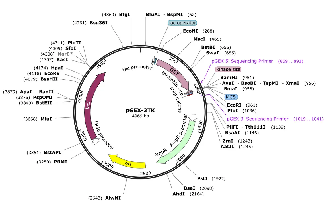
- Generate forward and reverse primers from this 1,353 nt sequence including: BamHI and EcoRI restriction sites to match with pGEX-2TK plasmid vector recipient (Figure 2)
- BamHI is 5'-GGATCC-3' and EcoRI is 5′-CCCGGG-3′
| BamHI restriction recognition site | EcoRI restriction recognition site |
|---|---|
| 5′ – G ↓G A T C C – 3′ | 5' – G ↓ A A T T C – 3' |
| 3′ – C C T A G ↑G – 5’ | 3' – C T T A A ↑ G – 5' |
- After designing and synthesizing forward and reverse primers that complement WRN500-946 1,353 nt and contain BamHI and EcoRI nt sequence, generate DNA copies of the fused gene through PCR
- Digest both PCR Product (BamHI-WRN500-946-EcoRI DNA sequence) and recipient plasmid MCS.
- Purify both DNA fragments by gel electrophoresis. Excise the desired bands and purify to remove contaminants that would inhibit ligation.
- Purify both DNA fragments by gel electrophoresis. Excise the desired bands and purify to remove contaminants that would inhibit ligation.
II. Clone and express GST-WRN500-946 plasmid vector
- Clone (plasmid vector containing WRN500-946 DNA insert) fused gene into Strain Bacteria Escherichia coli BL21 (DE3) pLysE translate genes to proteins to overpression of target protein by:
- Transform and start culture
- 9.1 Transform: Chemically competent BL21(DE3) with pGEX WRN plasmid vector
- 9.2 Plate: Inoculate 1:100 LB agar + ampicillin (100 µg/mL).
- 9.3 Incubate: 16 h at 37 °C, 200 rpm, to OD₆₀₀ = 0.5–0.6
- Inoculate:
- 10.1 Pick 3–4 colonies into 5 mL LB + amp.
- 10.2 Grow 37 °C, 200 rpm, to OD₆₀₀ ≈ 0.6–0.9
- Induce
- 11.1 Add IPTG (Isopropyl-1-thio-b-D-galactopyranoside) with 0.2 mM. Overnight (12h – 18h) at 16 °C (lowering temps to improve solubility and folding) . WRN500–946 typically expresses well at low temperature and remains soluble
- Harvest & Lysis [Perform on all remaining cells to get Large scale expression]
- 12.1. Prepare Lysis buffer
- 12.1.1. 50 mM Tris HCl, pH 7.5–8.0
- 12.1.2. 150 mM NaCl (can go 150–300 mM)
- 12.1.3. 1 mM DTT
- 12.1.4. 1 mM EDTA
- 12.1.5. 5–10% glycerol
- 12.1.6. 0.1–1% Triton X 100 or NP 40 (optional, helps solubility)
- 12.1.7. Protease inhibitors (EDTA free cocktail + 1 mM PMSF)
- 12.2. Procedure:
- 12.2.1. Transfer cell culture solution into centrifuge tube(s) and centrifuge at 10,000 rpm
- 12.2.2. Separate the solution by removing supernatant from cell pellet
- 12.2.3. Resuspend pellet: 5–10 mL lysis buffer per gram wet cell pellet.
- 12.2.3.1. Lyse: Sonication on ice (e.g., 10–15 cycles of 10 s on / 20 s off) Or high pressure homogenizer.
- 12.2.3.2. Clarify: Clear the lysate by centrifuge Spin 30 min at 15,000–20,000 g, 4 °C. if viscous, Filter supernatant through 0.45 µm.
III. Purification of GST-WRN500-946 helicase protein
- Prepare 1: Glutathione Resin e.g. Glutathione Sepharose 4B / 4 Fast Flow (Cytiva) or equivalent.
- Prepare 2: Equilibration / wash buffer: Same as lysis buffer, but you can drop detergent to 0–0.1% if needed.
- Binding step:
- 15.5 Equilibrate resin: 5–10 column volumes (CV) of cold buffer.
- 15.6 Bind: Add clarified lysate to resin (batch) or load onto a gravity column. Typical: 0.5–1 mL resin per liter culture. Incubate 1 h at 4 °C with gentle mixing rotation
- Wash: Wash with 10–20 CV of buffer until A₂₈₀ baseline stabilizes. For higher stringency, include: 300–500 mM NaCl, 0.1% Triton X 100, Optional: 5 mM MgCl₂ + 1 mM ATP to remove chaperones.
- Elution step:
- 17.1 Elution buffer: 50 mM Tris HCl, pH 8.0, 150 mM NaCl, 10 mM reduced glutathione, 5% glycerol, 1 mM DTT
- 17.2 Elute in 0.5–1 mL fractions, keep on ice.
- 17.3 Monitor A₂₈₀ and analyze fractions by SDS PAGE.
- Optional: Remove GST Tag. If you want tag free WRN fragment, include a thrombin or PreScission site in the construct and cleave on column or in solution.
IV. Polishing of GST-WRN500-946 helicase protein
- Size exclusion chromatography (SEC):
- Buffer: 20–25 mM Tris HCl, pH 7.5–8.0, 150 mM NaCl, 1 mM DTT, 5–10% glycerol
- Use Superdex 200 Increase 10/300 GL or similar
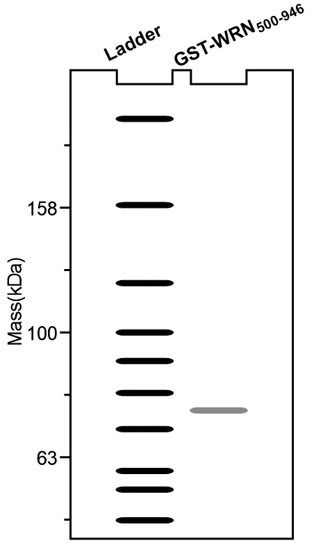
- Load 0.5–1 mL, run at 0.5–0.8 mL/min, collect 0.5 mL fractions. Pool monodisperse peak. You should see a single monodisperse peak around the expected size for GST fusion (~75 kDa). check by SDS PAGE
V. Storage of GST-WRN500-946 helicase protein
- Short term (days–weeks): 4 °C in SEC buffer + 10% glycerol, 1 mM DTT, 0.02% NaN₃.
- Long term (months): Aliquot, snap freeze in liquid N₂, store at −80 °C in: 20–25 mM Tris HCl (pH 7.5),150 mM NaCl, 1 mM DTT, 10–20% glycerol, Avoid repeated freeze–thaw cycles.
VI. Analysis of purified of GST-WRN500-946 Helicase protein
Spectrophotometer Absorbance A260/280
- WRN fragments commonly bind to DNA, which may require a check of the absorbance measurement to observe the ratio between peaks at 260nm and 280nm. A peak at 260nm indicates DNA impurities in the purified GST-WRN500-946.
Bradford Assay: Quantify concentrations GST-WRN500-947
- Materials: GST-WRN500-946, 5x Bradford reagent, bovine serum albumin (BSA) as a protein standard for calibration, and 1x PBS
- Instruments: centrifuge, plate reader or UV/Vis spectrophotometer, 96 well plate or standard quartz cuvettes and micropipettes
- General Instruction Protocol:
- Prepare 1X Working Solution of the Bradford reagent
- Prepare BSA protein standards solutions at appropriate concentrations based on the expected sample concentration
- Add 100 µL of 1X working solution into each well that contains standard, blank and sample solutions
- Shake to mix and incubate for 5 minutes at room temperature. The resulting signal is stable for 1 hour
- Measure the Optical Density (OD) at 595 nm
- Measurement: subtract the blank OD from the standard and sample absorbance. Plot the standards OD at 595 nm (y-axis) against the BSA concentration µg/mL (x-axis)
- Use the standard curve to calculate and determine the sample's protein concentration
SDS-PAGE and Coomassie staining
- Materials: GST-WRN500-946, purchased or handcast polyacrylamide gels, Prestained protein ladder (Invitrogen BenchMark Protein Ladder), Coomassie stain solution
- Instruments: electrophoresis chamber, micropipettes, spatula
- General Instruction Protocol:
- Perform the SDS-PAGE run by first preparing the sample and running buffers
- Prepare the gel(s) and assemble the electrophoresis chamber
- Prepare and load samples and ladder with diluted sample buffer at appropriate concentrations
- Perform the electrophoresis with run conditions and times based on the gel and buffer composition. The run times depend on the time required for the ladder’s dye front to reach the bottom of the gel cassette
- Analyze the protein separation by performing a total protein stain with Coomassie Staining Solution (e.g. Coomassie Brilliant Blue R-250 staining solution)
- Stain gels for 1–2 hours with gentle agitation. Stain overnight or longer if needed.
- Destain gel for 2 h. Change destaining solution multiple times (e.g., 3 washes x 30 min) until the background is less dark (figure 3).
VII. Expected results
- Yield: 4mg/L
- Purity: After GST affinity: ~70–85% and after SEC: >95%
- Chemical and Physical Properties:
- Monomeric
- Stable for months at −80 °C
Troubleshooting
| Problem | Possible Cause | Solutions/steps |
|---|---|---|
| Protein precipitation | High salt concentration | Slowly dialyze against buffer with lower salt |
| Low yield | Incomplete expression | Optimize induction temperature or media |
| Aggregation | Unstable folding | Add stabilizing agents or change pH |
References
- von Kobbe, C.; Karmakar, P.; Dawut, L.; Opresko, P.; Zeng, X.; Brosh, R. M., Jr.; Hickson, I. D.; Bohr, V. A. Colocalization, physical, and functional interaction between Werner and Bloom syndrome proteins. J Biol Chem 2002, 277 (24), 22035-22044.
- von Kobbe, C.; Thomä, N. H.; Czyzewski, B. K.; Pavletich, N. P.; Bohr, V. A. Werner syndrome protein contains three structure-specific DNA binding domains. J Biol Chem 2003, 278 (52), 52997-53006.
- Brosh, R. M., Jr.; von Kobbe, C.; Sommers, J. A.; Karmakar, P.; Opresko, P. L.; Piotrowski, J.; Dianova, I.; Dianov, G. L.; Bohr, V. A. Werner syndrome protein interacts with human flap endonuclease 1 and stimulates its cleavage activity. EMBO J. 2001, 20 (20), 5791-5801.
- Saydam, N.; Kanagaraj, R.; Dietschy, T.; Garcia, P. L.; Peña-Diaz, J.; Shevelev, I.; Stagljar, I.; Janscak, P. Physical and functional interactions between Werner syndrome helicase and mismatch-repair initiation factors. Nucleic Acids Res. 2007, 35 (17), 5706-5716.
- Muftuoglu, M.; Kulikowicz, T.; Beck, G.; Lee, J. W.; Piotrowski, J.; Bohr, V. A. Intrinsic ssDNA annealing activity in the C-terminal region of WRN. Biochemistry 2008, 47 (39), 10247-10254.
- Lee, J. W.; Kusumoto, R.; Doherty, K. M.; Lin, G. X.; Zeng, W.; Cheng, W. H.; von Kobbe, C.; Brosh, R. M., Jr.; Hu, J. S.; Bohr, V. A. Modulation of Werner syndrome protein function by a single mutation in the conserved RecQ domain. J Biol Chem 2005, 280 (47), 39627-39636.
- Harrigan, J. A.; Opresko, P. L.; von Kobbe, C.; Kedar, P. S.; Prasad, R.; Wilson, S. H.; Bohr, V. A. The Werner syndrome protein stimulates DNA polymerase beta strand displacement synthesis via its helicase activity. J Biol Chem 2003, 278 (25), 22686-22695.